ProfessionalsMembersResourcesDownloadsGuidelinesParasite ID PostersParasite Life CyclesPodcastsTherapiesTick Hosts, Habitats and PreventionFAQsScientificControlEctoparasitesEndoparasitesVector Borne DiseasesZoonosesTravelling Pets
There are a wide range of helminths (worms) that can infect dogs and cats, including:
Major helminth groups by location in the host are:
Intestinal Worms

Cestodes:
Nematodes:
Non-Intestinal Worms
Nematodes
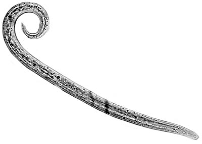
Nematodes are the most common worm in both dogs and cats with a high prevalence in the UK and throughout Europe. They are circular in cross section and have openings at both ends. They are found in either the gastrointestinal tract, the respiratory tract or the heart. A number of roundworm species are also categorised under further helminth groups, for example hookworms, whipworms, heartworms and lungworms.
Factors affecting the importance of these helminths include:
Use the links above to see more information about each type of helminth
Helminth management requires a customised combination of management and anthelmintic treatment suitable for the individual animal or animals. Anthelmintic treatments should be repeated at suitable intervals and should cover the spectrum of helminths present or likely to be present in the pet. Where preferred, anthelmintic treatments can be replaced by faecal examination conducted regularly, with subsequent anthelmintic treatment where indicated by a positive result.
Ascarids, are a type of nematode (also sometimes referred to as roundworms) more commonly found in young, stray or unneutered animals. Most animals are susceptible at any age. Animals are commonly infected through hunting or ingesting worm eggs in the environment. A number of species also have zoonotic potential and so additional measures should be taken to reduce risks to human health.


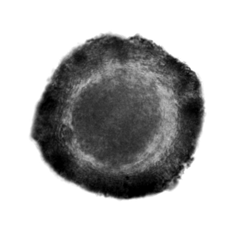
Toxocara spp. (T. canis in dogs and T. cati in cats)
Parasite type: Nematode; Ascarid
Location: Small intestine
Key notes: These parasites are prolific egg-layers and just a few worms can produce large numbers of eggs. Toxocara spp. eggs can survive in the environment for months or even years. High risk of prenatal infection in dogs.
Transmission: Ingestion of larvae via predation of paratenic hosts (e.g. rodents) or infective eggs from the environment.
Puppies can be heavily infected by T. canis worms in utero and both kittens and puppies via nursing. These may cause serious illness before diagnosis is possible by faecal examination.
Prepatent period: 4-5 weeks following ingestion of eggs or larvae in a paratenic host.
Clinical signs: Often appear normal with a light infection. A heavy infection may cause cachexia and a pot-bellied appearance. Worms may be passed in the faeces or in vomit. Migrating larvae through the lungs can cause verminous pneumonia associated with coughing. Retinal dysfunction may occur in dogs due to migrating larvae; although it does not usually affect the animal's normal functioning, lesions may be seen on examination of the retina.
In kittens and pups: respiratory signs, cachexia, gastrointestinal dysfunction.
Diagnosis: Modified McMaster technique for eggs.
Control: Management measures such as feeding cooked food, hygiene including removal and disposal of faeces will assist in reducing the risk of infection.
For puppies and kittens
Puppies should be treated with an appropriate anthelmintic (see therapy tables) beginning at the age of two weeks. Subsequently the treatment should be repeated fortnightly until two weeks after weaning.
Kittens should be treated with an adequate anthelmintic (see therapy tables) beginning at the age of three weeks on the assumption that the queen is infected with Toxocara cati. According to present experience a fortnightly treatment until two weeks after weaning is advisable.
Nursing bitches and queens should be treated concurrently with the first treatment of their offspring as they often develop patent infections at this time.
Nursing bitches and queens
Nursing bitches and queens should be treated concurrently with the first treatment of their offspring as they often develop patent infections at this time.
For adult dogs and cats
Current information suggests annual or twice yearly treatments do not have a significant impact on preventing patent infection within a population, so a treatment frequency of at least 4 times per year is a general recommendation.
Studies have shown that worming four times a year does not necessarily eliminate patent infections, while a monthly worm treatment can largely prevent patent infections as it takes into account the biology of the parasites.
In cases of increased risk (such as kennels or households where there are children) monthly treatment can minimise the risk of patent infections and the excretion of infective parasite stages, as the prepatent period for Toxocara spp. is a little more than four weeks.
Toxascaris leonina
Parasite type: Nematode; Ascarid
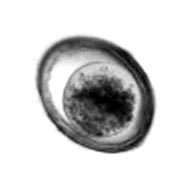
Location: Small intestine
Key notes: Zoonotic. Rodents act as intermediate hosts.
Transmission: Cats and dogs become infected by ingestion of embryonated egg or infected rodent. There is no prenatal or lactogenic transmission.
Prepatent period: 12 weeks for cats and 8 weeks for dogs
Clinical signs: normally no clinical signs
Diagnosis: Modified McMaster technique
Control: Management measures such as feeding cooked food, hygiene including removal and disposal of faeces will assist in reducing the risk of infection. Regular worming to control Toxocara spp. will also control T. leonina so long as a treatment with an appropriate spectrum of activity is selected.
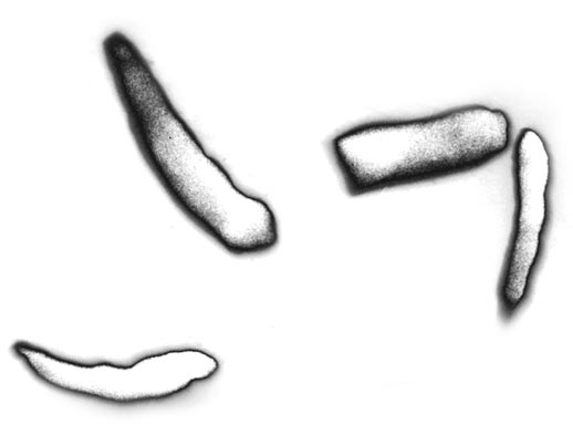
Strongyloides spp. (threadworm)
Parasite type: GI Nematode; Ascarid
Location: Small intestine
Key notes: Largely found in puppies. Infection can be passed to puppies whilst nursing.
Transmission: Larvae penetrate the skin and travel through the blood stream to the lungs of the host. The larvae are then coughed up and swallowed.
Prepatent period: 1 week
Clinical signs: normally no clinical signs
Diagnosis: Modified McMaster technique or other flotation technique
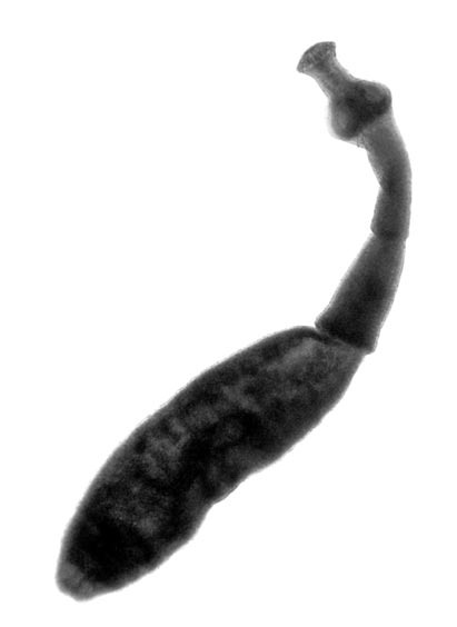
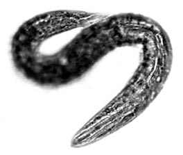
Hookworms
Hookworms are small nematodes characterised by large mouthparts that are at an angle to the rest of the worm, hence the common name. There are three species of significance in Europe: Ancylostoma caninum, Ancylostoma tubaeforme and Uncinaria stenocephala. In the UK U. stenocephala is the most common, with A. caninum seen less frequently.
Ancylostoma caninum
Parasite type: GI Nematode; Hookworm
Location: Small intestine
Key notes: A degree of immunity develops after infection. Infection can be passed to puppies when nursing.
Transmission: Adult worms have a direct life cycle with eggs passed in the faeces developing to third stage larvae (L3) in the environment. When these are ingested they develop within 2-3 weeks to adult worms. Ancylostoma spp. larvae are capable of penetrating skin and thus making their way to the intestine.
Prepatent period: 2-3 weeks
Clinical signs: Anaemia in a heavy infection
Diagnosis: Modified McMaster Technique
Control: Prompt removal of faeces and disposal will help to prevent larvae developing on grass. Regular worming to control Toxocara spp. will also control hookworms so long as a treatment with an appropriate spectrum of activity is selected.
Ancylostoma tubaeforme
Parasite type: GI Nematode; Hookworm
Location: Small intestine
Key notes: A degree of immunity develops after infection. Infection can be passed to kittens when nursing.
Transmission: Adult worms have a direct life cycle with eggs passed in the faeces developing to third stage larvae (L3) in the environment. When these are ingested they develop within 2 – 3 weeks to adult worms. Ancylostoma spp. larvae, are capable of penetrating skin and thus making their way to the intestine.
Prepatent period: 2-3 weeks
Clinical signs: Anaemia
Diagnosis: Modified McMaster Technique
Uncinaria stenocephala (Northern Hookworm)

Parasite type: GI Nematode; Hookworm
Location: Small intestine
Key notes: Also known as Northern Hookworm. A degree of immunity develops after infection.
Transmission: Adult worms have a direct life cycle with eggs passed in the faeces developing to third stage larvae (L3) in the environment. When these are ingested they develop within 2 – 3 weeks to adult worms.
Prepatent period: 2-3 weeks
Clinical signs: Normally no clinical signs
Diagnosis: Modified McMaster Technique
Control: Prompt removal of faeces and disposal will help to prevent larvae developing on grass. Regular worming to control Toxocara spp. will also control hookworms so long as a treatment with an appropriate spectrum of activity is selected.
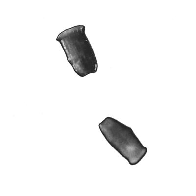
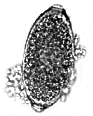
Whipworms
Trichuris vulpis
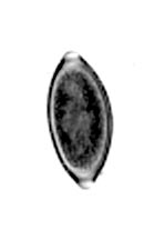
Parasite type: GI Nematode; Whipworm
Location: Large intestine
Key notes: Considerable and persistent contamination of the environment can occur. Infection can be diagnosed by finding characteristic “lemon-shaped” eggs in faeces.
Transmission: Eggs are passed in the faeces of infected dogs and the first stage larva (L1) develops within the egg in 1-2 months. No development occurs below 4°C. The larvae are protected by the egg shell and can survive in the environment for years. Dogs are infected when they eat eggs containing infective larvae (L1). The pre-patent period is 2 - 3 months and infected dogs may continue to shed eggs for up to a year.
Prepatent period: 8-12 weeks
Clinical signs: A light infection is well tolerated. A heavy infection will result in diarrhoeic bloody, mucus-filled faeces and ultimately, the animal will no longer be able to compensate and will become acutely ill. Metabolic disturbance including hyponatraemia may be seen.
Diagnosis: Modified McMaster technique
Control: Control depends on removing dogs where possible from the contaminated environment and repeated anthelmintic treatment. Since the eggs are difficult to eliminate from the environment it may be necessary to pave or plough affected areas. Prompt removal of faeces and disposal will help to prevent eggs developing in the environment.
Regular worming to control Toxocara spp. will also control hookworms so long as a treatment with an appropriate spectrum of activity is selected.
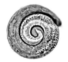
Tapeworms are cestodes which live in the small intestine. They consist of a chain of proglottids (segments), which attach to the intestinal wall of the animal with a hold-fasting organ known as a scolex. When mature, tapeworms shed distal segments containing eggs which are passed in the faeces into the environment.
Tapeworms have an indirect lifecycle requiring an intermediate host where the larval stages develop. Larval or metacestodes usually take the form of cysts within the tissues of the intermediate hosts and primary control measures therefore include preventing exposure to intermediate hosts, where possible.
Tapeworms have a high prevalence across Europe, including a number of zoonotic species which pose a serious health risk to humans.
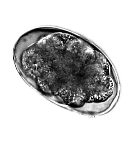
Echinococcus granulosus
Parasite type: Cestode; Tapeworm
Location: Small intestine
Key notes: Zoonotic – causes Hydatid Disease in humans.
Transmission: Intermediate hosts, primarily sheep but also cattle and horses, become infected when they ingest eggs from the environment, resulting in the formation of hydatid cysts in the liver and lungs. Infection results in dogs when they ingest material containing these hydatid cysts. Eggs passed in the faeces of the final hosts are immediately infective to intermediate hosts including humans.
Prepatent period: 5-8 weeks
Clinical signs: No clinical signs in the canine final host.
Diagnosis: Modified McMaster technique or segment identification. Specific diagnosis of Echinococcus spp. infections in definitive hosts is difficult as the taeniid-eggs cannot be differentiated morphologically and are passed intermittently. In Echinococcus spp. endemic areas taeniid-infections based on egg detection should be handled as potential Echinococcus spp. infection. Where animals are infected with Echinococcus species, it is advisable that they are treated under the supervision of a veterinarian on two consecutive days with a highly effective anthelmintic and that the dogs are shampooed to remove any parasite eggs adhering to the coat. The personnel involved should use suitable protective clothing such as protective gloves and a mask.
Control: Care should be taken to prevent dogs having access to raw offal and carcasses. Additionally, in areas where E. granulosus is endemic (Ovine and Equine species in the UK and ovine, equine, porcine and bovine species in Europe) is endemic, dogs that may have access to carcasses or raw viscera should be treated at least every 6 weeks with an effective anthelmintic containing praziquantel or epsiprantel.
Echinococcus multilocularis
Parasite type: Cestode; Tapeworm
Location: Small intestine
Key notes: Zoonotic – causes alveolar echinococcosis in humans. Foxes are the main definitive hosts and voles act as intermediate hosts. Infection uncommon but not impossible in cats. The mandatory tapeworm treatment within the PETs scheme prior to entry into the UK is designed to prevent its entry.
Transmission: Eggs are infective immediately when passed into the environment. Intermediate hosts (small rodents and voles) become infected when they ingest eggs from the environment and this results in the development of alveolar cysts in the liver (and occasionally other organs). Infection results in the definitive hosts (primarily foxes and dogs) when they ingest material containing these cysts. Protoscolices contained within the cysts develop into tapeworms in the definitive host, resulting in eggs being passed in the faeces which are immediately infective to intermediate hosts, including humans.
Prepatent period: 4-8 weeks
Clinical signs: No clinical signs in the canine or feline host.
Diagnosis: Modified McMaster technique or segment identification. Specific diagnosis of Echinococcus spp. infections in definitive hosts is difficult as the taeniid-eggs cannot be differentiated morphologically and are passed intermittently. In Echinococcus spp. endemic areas taeniid-infections based on egg detection should be handled as potential Echinococcus spp. infection.
Control: In endemic areas, dogs that have access to rodents should be treated at four weekly intervals with an effective anthelmintic containing praziquantel. Cats, in contrast to dogs, are epidemiologically insignificant as sources of egg output as they are poor hosts for this worm, although they do sporadically acquire infection and occasionally pass eggs. In contrast to dogs, where it is common to find eggs in the fur of infected animals, no eggs have been recovered to date from the coat of an infected cat. Since there is a small risk of cats carrying an infection, it is reasonable to recommend treatment in high risk situations, for example prior to entry into countries where the infection is not present.
Where animals are infected with Echinococcus species, it is advisable that they are treated under the supervision of a veterinarian on two consecutive days with a highly effective anthelmintic and that the dogs are shampooed to remove any parasite eggs adhering to the coat.
The personnel involved should use suitable protective clothing such as protective gloves and a mask.
Dipylidium caninum
Parasite type: Cestode; tapeworm
Location: Small intestine
Key notes: Zoonotic, VBD (although infection is unwelcome, it is unlikely to cause severe illness in a human).
Transmission: This tapeworm of dogs or cats has the flea or the chewing louse as its intermediate host. Infection occurs as a result of ingesting infected insects.
Prepatent period: 3 weeks
Clinical signs: rarely associated with clinical signs in the dog or cat. Motile segments.
Diagnosis: Segment identification or Modified McMaster technique.
Control: Preventative measures include controlling flea and lice infestations.
Dogs or cats can be treated with an appropriate cestocide to remove infection. Unless flea and/or louse infestations are controlled, reinfection can occur with signs of infection present after as little as three weeks.
Taenia spp.
Parasite type: Cestode; tapeworm
Location: Small intestine
Key notes: Patency can last for months or years.
Transmission: Taenia taeniaeformis, the species that occurs in cats, uses rodents as intermediate hosts. Dogs or cats are infected when they eat tissues or viscera of infected hosts.
Prepatent period: The prepatent period for Taenia spp. ranges from about 4 - 10 weeks in dogs (depending on species) and is approximately 5 - 10 weeks for Taenia taeniaeformis in cats.
Clinical signs: Well tolerated. Anal irritation, motile segments.
Diagnosis: Segment identification or Modified McMaster technique.
Control: Preventative measures include deterring hunting. Dogs or cats can be treated with an appropriate cestocide to remove infection. Unless hunting is controlled, reinfection can occur.
Mesocestoides spp.
Parasite type: Cestode; tapeworm
Location: Small intestine
Key notes: A wide range of animals can act as intermediate hosts, including birds, reptiles, amphibians and mammals.
Transmission: Intermediate hosts are infected when they ingest infected mites or insects which have ingested eggs from the environment. The metacestode stage develops in the intermediate host and infection is passed to definitive hosts (foxes, dogs and occasionally cats) when they ingest an infected animal. Eggs are then passed in the hosts’ faeces into the environment.
Prepatent period: 3-4 weeks
Clinical signs: No clinical signs
Diagnosis: Modified McMaster technique or centrifugal flotation techniques.
Control: Preventative measures include deterring hunting.
Heartworms are nematodes whose adult stages occur in the heart or vasculature. Despite residing in the circulatory system, clinical signs often include respiratory problems due to lung pathology. Heartworms are prevalent in Southern Europe and are also found in the UK.
Dirofilaria immitis
Parasite type: Nematode; Heartworm
Location: Pulmonary artery, occasionally found in the right heart and adjacent large vessels such as the cranial and caudal venae cavae. Ectopic localizations in the brain, eyes, testis or aorta occur rarely and more especially in cats.
Key notes: Also known as ‘Heartworm’. Zoonotic. VBD. Dogs are the primary definitive host. The cat is a susceptible, but not ideal, host. D. immitisis endemic in Mediterranean areas. Individual cases may be seen in the UK where dogs or cats have returned from Mediterranean or other endemic areas. There are believed to be mosquitoes capable of supporting the development of D. immitis, therefore it is critical that dogs and cats travelling to endemic areas receive appropriate heartworm prevention in order to prevent the introduction and establishment of the infection in the UK.
Transmission: D. immitis microfilariae develop in the uterus of female worms and are passed into the blood stream where they become available to blood-sucking mosquitoes. Microfilariae develop to the infective stage (L3) in the body of these vectors and are transmitted during feeding. Larvae undertake an extensive migration through subcutaneous, subserosal and muscular tissues to reach the pulmonary arteries and the right heart where they develop to the adult stage and mate. In dogs, adult worms are able to survive up to 7 years.
Many mosquito species are competent intermediate hosts and allow the development of microfilariae to infective stages. The most important vectors in Europe are species of the genera Culex, Aedes, and Anopheles. Recently, Aedes albopictus, the Asian Tiger Mosquito which is currently spreading in Europe, has been shown to be a competent vector for Dirofilaria spp. under field conditions.
Prepatent period: 6-7 months
Clinical signs: The clinical evolution of heartworm disease in dogs is usually chronic. Most infected dogs do not show any clinical signs for years depending on the worm burden, individual sensitivity and level of exercise; arterial damage is usually more severe in dogs that perform intensive physical exercise. Clinical signs of the disease develop gradually and may begin with a chronic cough that may be followed by moderate to severe dyspnoea, weakness, and sometimes syncope after exercise or excitement. At this stage, auscultation may reveal abnormal pulmonary sounds (crackles) over the caudal lung lobes and a split second heart sound can often be heard. Later, when right congestive heart failure is developing, oedema of the abdomen and less often the limbs may be observed together with anorexia, weight loss and dehydration. Cardiac murmurs over the right side of the thorax due to tricuspid valve insufficiency and abnormal cardiac rhythm due to atrial fibrillation are common findings. Sudden death is rare and usually occurs following respiratory distress or progressive emaciation. During the chronic stages of the disease, there may be a sudden onset of acute signs. For example, after severe spontaneous thromboembolism following the natural death of many heartworms, dogs may show acute life-threatening dyspnoea and haemoptysis.
In small dogs, the displacement of adult worms from the pulmonary arteries to the right heart due to pulmonary hypertension and a sudden fall in right cardiac output, is a common event. In this case, affected dogs present the so-called “caval syndrome”. Dyspnoea, a tricuspid cardiac murmur and haemoglobinuria, due to mechanical haemolysis in the right cardiac chambers, are the most typical signs and the outcome is usually fatal.
Clinical signs in cats are quite different from those in dogs. Most cats show no clinical signs for a long time after infection. These cats may undergo spontaneous self-cure due to the natural death of parasites without showing any signs or they may suddenly show a dramatic acute syndrome where respiratory signs such as coughing, dyspnoea and haemoptysis are usually seen; vomiting also occurs frequently. Sudden death in apparently healthy cats is not an infrequent consequence of infection. Chronic illness including coughing, vomiting, diarrhoea and weight loss is less frequently observed. In contrast to the situation in dogs, clinical signs related to right ventricular heart failure are not considered consistent with heartworm infection in cats. In most cases, the onset of clinical signs seems to be related to the natural death of parasites or to the arrival of pre-adult heartworms (L5) in the pulmonary arteries.
Diagnosis: Heartworm infection in dogs can be detected with blood tests which demonstrate the presence of circulating microfilariae (in wet blood smears or Knott test) or adult antigens (in whole blood, serum or plasma samples), but further diagnostic procedures are usually required to determine the severity of disease and possible treatment options. Morphological differentiation of the microfilariae is often difficult because of overlap in lengths of most species. However, microfilariae can be differentiated in specialised laboratories by the acid phosphatase stain (APh-S) or by molecular investigations (PCR).
Control: Currently there are no repellents/insecticides that have been demonstrated to prevent transmission of heartworm, therefore control in dogs and cats depends on the use of heartworm preventive treatments that kill the young heartworm stages prior to their migration towards the heart.
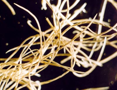
Angiostrongylus vasorum
Parasite type: Nematode; Heartworm
Location: Pulmonary arteries and right side of the heart.
Key notes: Commonly known as ‘Lungworm’ in the UK. Also known as French Heartworm. A. vasorum occurs in the UK and has been seen increasingly commonly over an increased geographic range (now including Scotland) over the past decade.
Transmission: Slugs and snails act as intermediate hosts. Dogs may also acquire infection through ingestion of frogs and other amphibians acting as paratenic hosts.
Following ingestion of L3 by a dog, larvae develop and migrate to the right side of the heart and pulmonary artery. Female worms begin laying eggs from 38-60 days after infection.
Eggs hatch rapidly and larvae penetrate the alveoli, are coughed up and passed in faeces as first stage larvae (L1). Once an infection is established, patency is very long – possibly lifelong if left untreated.
Prepatent period: 4-8 weeks
Clinical signs: In early infections, coughing (may be harsh and dry), dyspnoea, anaemia, depression, anorexia, and signs of coagulopathy e.g. melaena, haemoptysis, prolonged bleeding from minor injuries and subcutaneous haematomas may be seen. In severe infections right sided heart failure and even sudden death may occur.
In chronic infection, verminous pneumonia can develop leading to anorexia and weight loss, emaciation and pulmonary hypertension.
Occasionally larvae and rarely adult stages of A. vasorum are located in ectopic locations such as brain, bladder, kidney or the anterior chamber of the eye. This may result in clinical signs related to invasion of these organs.
Diagnosis: Baermann Technique for L1 larvae
Lab based and patient side antigen PCR blood testing
Control: Anthelmintic therapy includes use of macrocyclic lactone based anthelmintics or repeated daily administration of benzimidazole based anthelmintics (5 days to several weeks). Administration of macrocyclic lactone based products has been shown to be effective. Supportive treatment, with antibiotic and glucocorticoid based products may be needed in severe clinical cases and the animal should be rested during the treatment period (at least 2-3 days).
Prevention of disease requires prevention of access to mulluscs and amphibians and/or monthly treatment with an anthelmintic licensed for A. vasorum prevention.
Lungworms are nematodes whose adult stages occur in the respiratory tract of the host. There are many different lungworm parasites which occupy different areas within the respiratory system. Lungworms are prevalent throughout the UK and Europe and can be transmitted directly from host to host or via intermediate hosts, depending on the species.
Aelurostrongylus abstrusus
Parasite type: Nematode; Lungworm
Location: Lung parenchyma
Key notes: Also known as ‘Cat Lungworm’.
Transmission: Slugs and snails act as intermediate hosts. Cats may also acquire infection through ingestion of frogs and other amphibians acting as paratenic hosts.
Prepatent period: 4-6 weeks
Clinical signs: Mild infection may be unnoticeable. Coughing and sneezing may present as clinical signs. Dyspnoea may be present in severe infections.
Diagnosis: Baermann Technique
Control: Anthelmintic therapy. Preventative measures include preventing contact with amphibians and controlling gastropods within the cat’s territory.
Oslerus osleri
Parasite type: Nematode; Lungworm
Location: Trachea and bronchi
Key notes: Formerly Filaroides osleri
Transmission: Passes directly from host to host with no need for an intermediate host. Often passed from bitch to pups so may be present in a litter of pups by 10 weeks of age.
Prepatent period: 10 Weeks
Clinical signs: Mild infection may be unnoticeable. Exercise intolerance, coughing.
Diagnosis: Baermann technique
Control: Anthelmintic therapy
Filaroides hirthi
Parasite type: Nematode; Lungworm
Location: Lung parenchyma
Key notes: Not commonly found in the UK
Transmission: Passes directly from host.
Prepatent period: 32-35 days
Clinical signs: Cough which worsens through exercise
Diagnosis: Baermann techinique
Control: Use Anthelmintic treatment. Continue to faecal test after treatment to ensure that the infection is cured.
Crenosoma vulpis
Parasite type: Nematode; Lungworm
Location: Trachea and bronchi
Key notes: Also known as the Fox Lungworm
Transmission: Through ingesting a snail, slug or other paratenic host
Prepatent period: 3 Weeks
Clinical signs: Asymptomatic – cough, retching, reduced appetite
Diagnosis: Baermann technique
Control: Anthelmintic therapy
+/- preventing access to molluscs and amphibians
Capillaria aerophila
Parasite type: Nematode; Lungworm
Location: Trachea and bronchi
Key notes: Also known as Eucoleus aerophilus
Transmission: Passes directly from host to host.
Prepatent period: 6 weeks
Clinical signs: May be unnoticeable or slight respiratory signs
Diagnosis: Modified McMaster technique for characteristic eggs with bipolar plugs
Control: Anthelmintic therapy
Subcutaneous worms and eye worms
Some worms inhabit less common areas of the body, such as subcutaneous tissue and the eye. These worms are members of the roundworm family and are therefore circular in cross section and have openings at both ends. However, they are transmitted via vectors and do not migrate around the body of the final host. These worms are zoonotic and are prevalent in certain areas of Europe but not in the UK.
Dirofilaria repens
Parasite type: Nematode; Subcutaneous worm.
Location: Subcutaneous tissue, muscular fasciae.
Key notes: Also known as ‘Subcutaneous Worm’. Zoonotic. VBD. Occurs in many areas of central and southern Continental Europe with an increasing geographic range. The main concern relating to D. repens in Europe is its ability to cause human infection.
Transmission: D. repens microfilariae develop in the uterus of female worms and are excreted into the blood stream where they become available to blood-sucking mosquitoes. Microfilariae develop to the infective stage (L3) in the body of these vectors and are transmitted via their saliva during feeding. D. repens infective larvae do not migrate far in the subcutaneous connective tissues and reach maturity there. Adult worms are found between subcutaneous and deep connective tissue layers in most parts of the body, sometimes forming non-inflammatory nodules. Adults can live for several years.
Many mosquito species are competent intermediate hosts and allow the development of microfilariae to infective stages. Recently, Aedes albopictus, the Asian Tiger Mosquito which is currently spreading in Europe, has been shown to be a competent vector for Dirofilaria spp. under field conditions.
Prepatent period: 27-34 weeks.
Clinical signs: Disease due to D. repens infection is much less severe than that due to heartworm D. immitis infection and is frequently sub-clinical. However, infected dogs can present with cutaneous disorders of varying severity, such as pruritus, dermal swelling and subcutaneous nodules which contain the parasites. Severe disease has been associated with allergic reactions probably against microfilariae.
Diagnosis: D. repens infection in dogs can be detected with blood tests which demonstrate the presence of circulating microfilariae (in wet blood smears or Knott test) or adult antigens (in whole blood, serum or plasma samples), but further diagnostic procedures are usually required to determine the severity of disease and possible treatment options. Morphological differentiation of the microfilariae is often difficult because of overlap in lengths of most species. However, microfilariae can be differentiated in specialised laboratories by the acid phosphatase stain (APh-S) or by molecular investigations (PCR).
Control: Like heartworm infection, subcutaneous filariosis can be safely and effectively prevented in both dogs and cats by chemoprophylactic treatments. Monthly treatments with macrocyclic lactones (oral or spot-ons formulations) or an annual treatment with an injectable sustained release formulation at the same doses used against D. immitis, once at the beginning of the risk season, have been found to be effective in preventing subcutaneous infection in dogs naturally exposed to D. repens transmitting mosquitoes.
Thelazia callipaeda
Parasite type: Nematode; Eye worm.
Location: Surface of the cornea.
Key notes: VBD. Zoonotic.
Transmission: Eggs are released into the tears of the host. Tear feeding flies (Muscid flies) then ingest the eggs and act as intermediate hosts to the L1 larvae which penetrate the fly's intestinal wall and develop into L3 larvae. The larvae migrate to the head of the intermediate host and are released into or near the eye when the fly feeds. L3 larvae develop into adult worms in the hosts’ eyes and then lay eggs.
Prepatent period: 3 weeks.
Clinical signs: Epiphora, mucopurulent discharge, lacrimation, conjunctivitis, corneal opacity, corneal ulceration.
Diagnosis: Diagnosis can be made by identifying adult worms in the hosts’ eyes.