Also known as the Brown dog tick, kennel tick
Hosts
Most mammals: particularly dogs, humans, occasionally cattle)
Distribution
Worldwide
Rhipicephalus sanguineus is generally found in sheltered areas where climactic conditions remain relatively constant. Large concentrations of populations are found mainly in warm and moist environments.
Pathogenesis
Dog: Babesia canis, Babesia gibsoni, Ehrlichia canis
Human: Rickettsia rickettsi, Rickettsia sibirica, Rickettsia conori, Francisella tularensis
Other diseases that have been associated with R. sanguineus include:
Rickettsia rhipicephali
Haemobartonella canis
Hepatozoon canis
Dipetalonema dracunculoides
Description
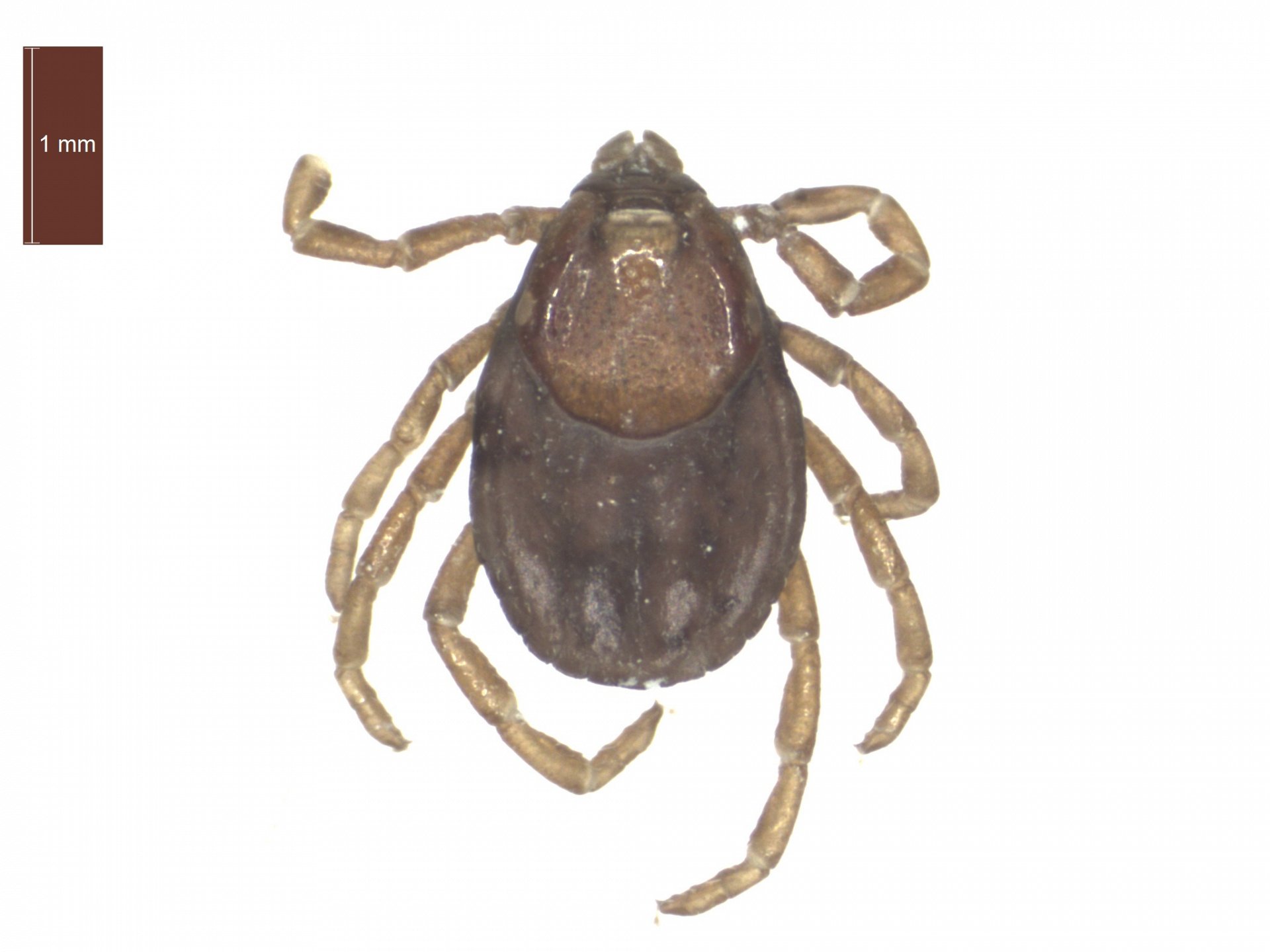
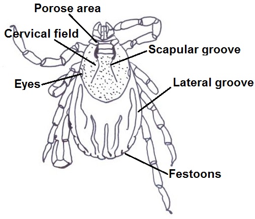
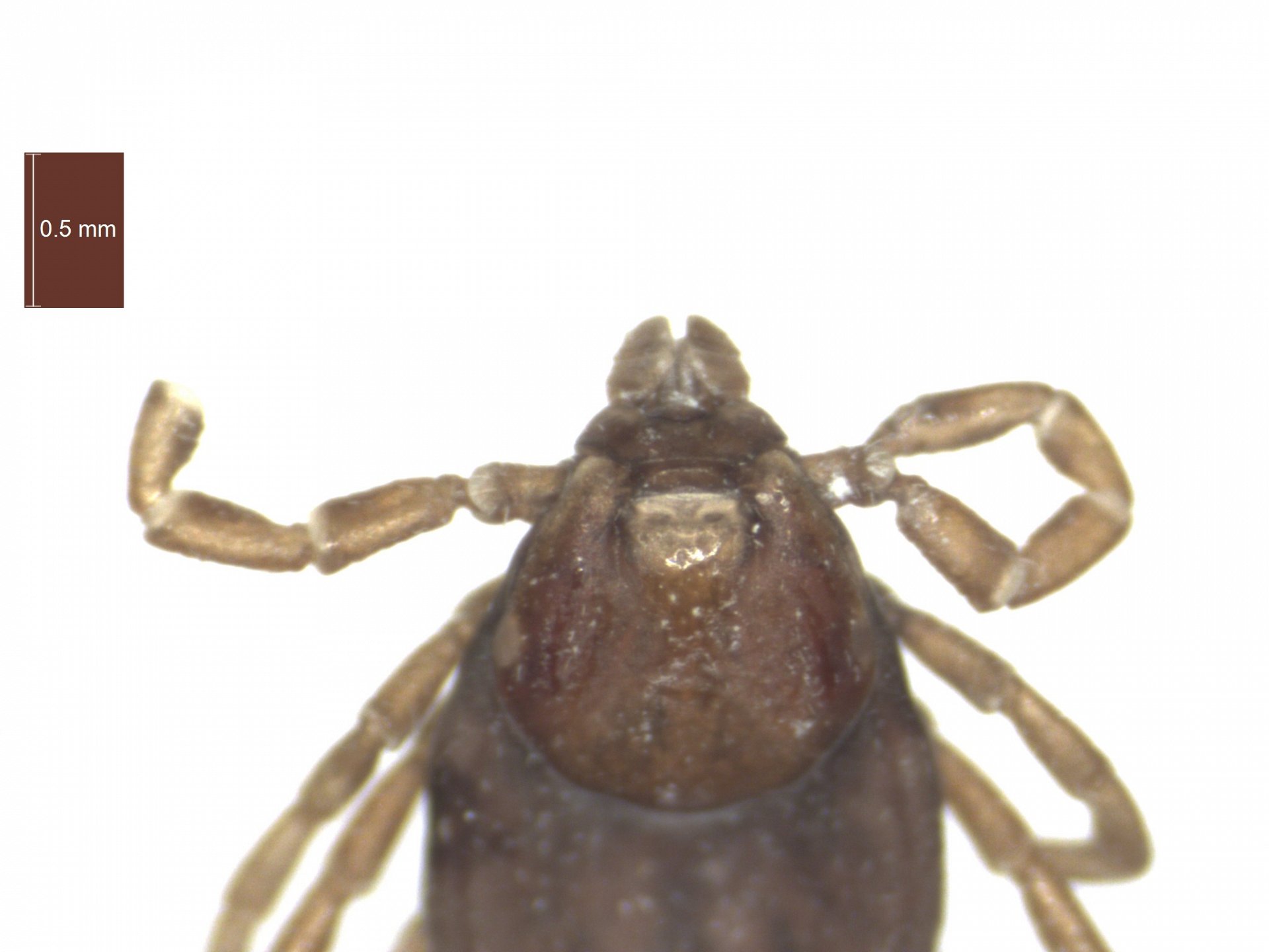
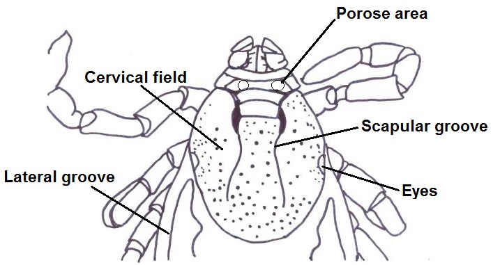
Adult female dorsal features
- Porose areas are circular and broadly separated
- Cervical field depressions are large and straight
- Scapular grooves are steep
- Scutum is pale in colour
- Unfed females can measure between 3.0mm-4.5mm in length
- Engorged females can measure up to 12mm in length
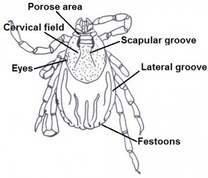
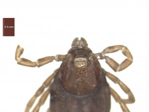

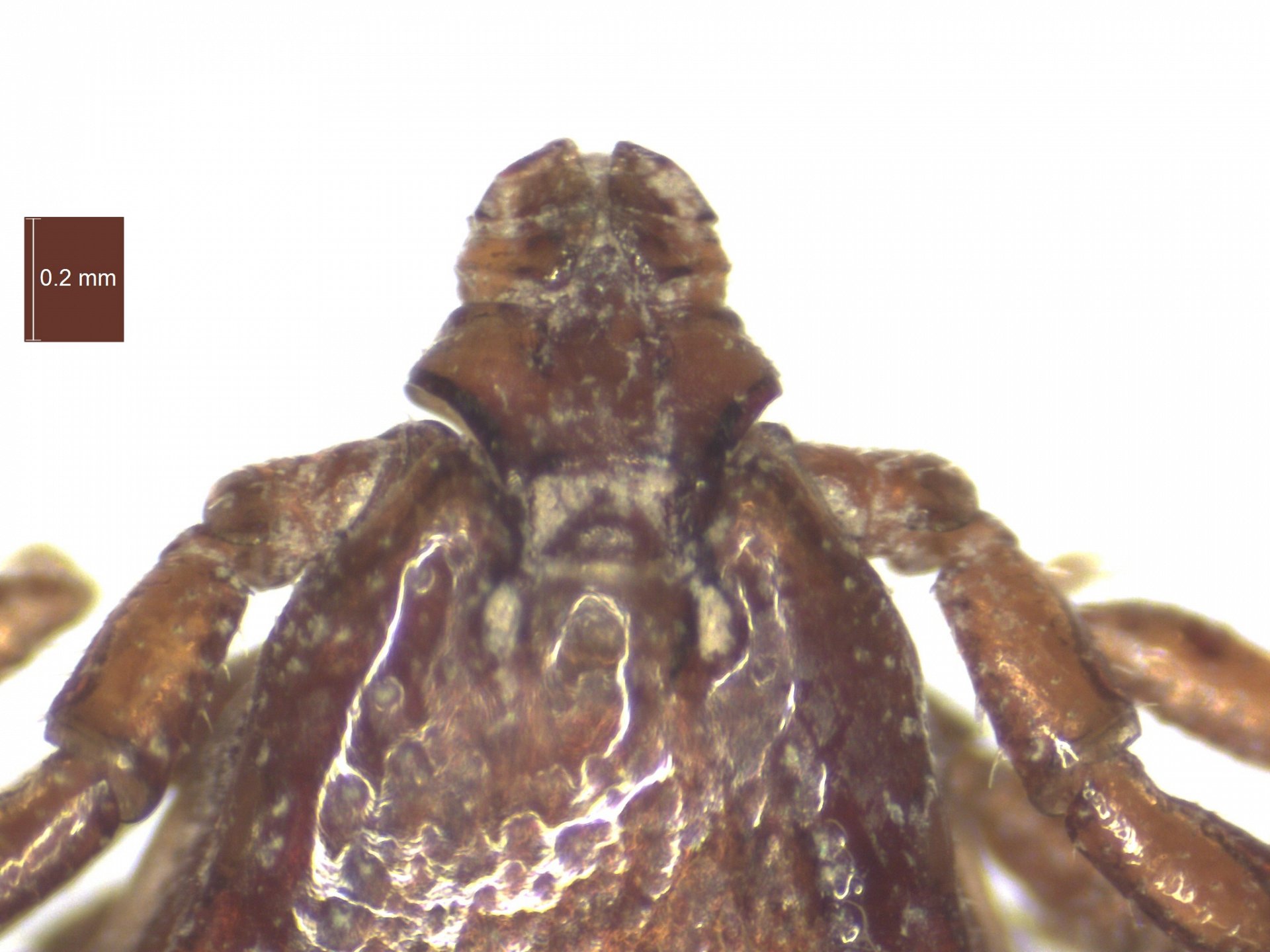
Adult female dorsal view and adult female close up of gnathosoma. (Click on pictures for a close up)
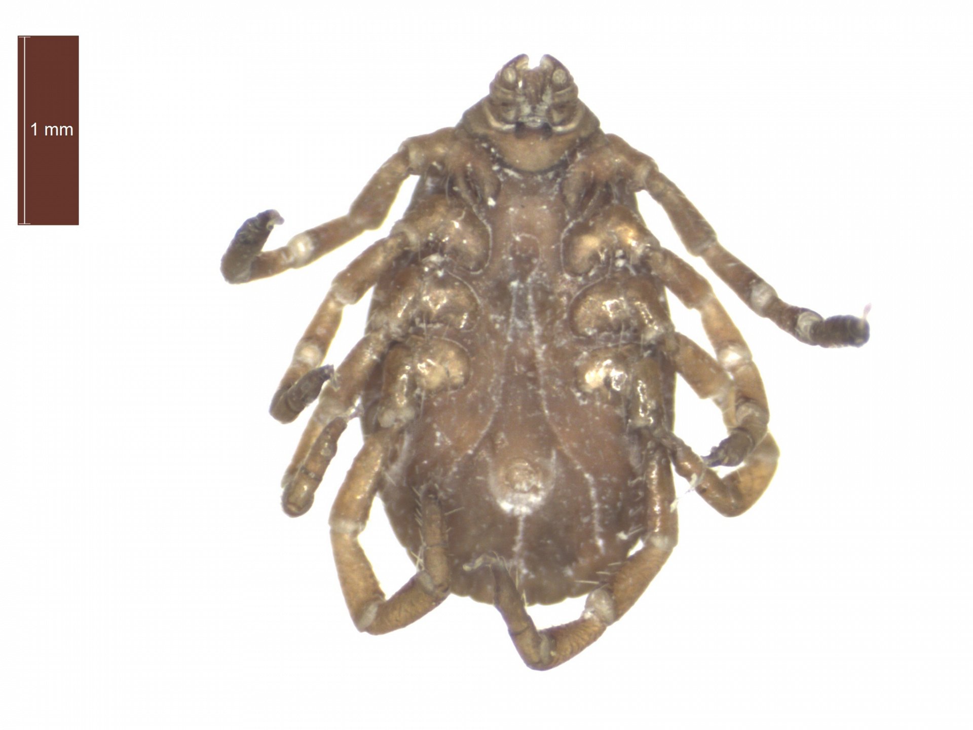
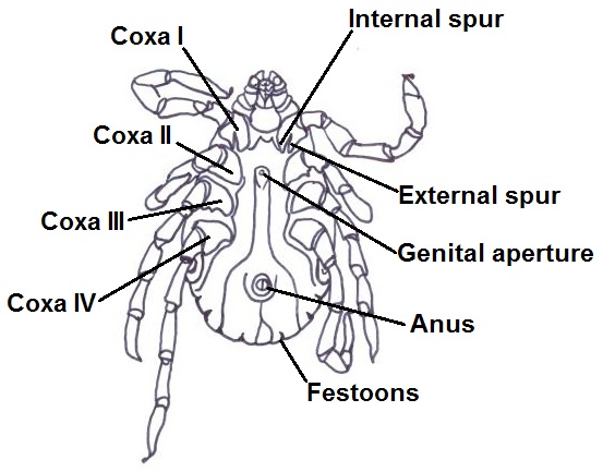
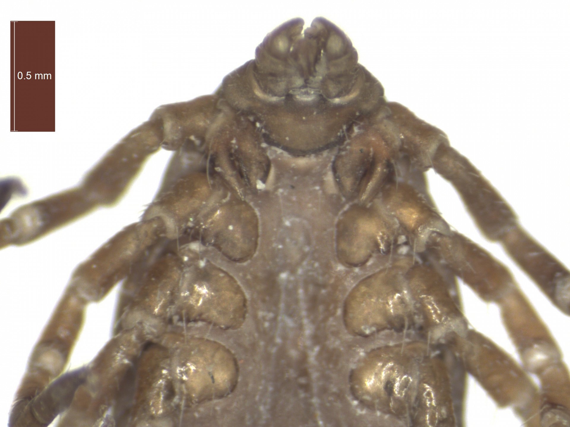
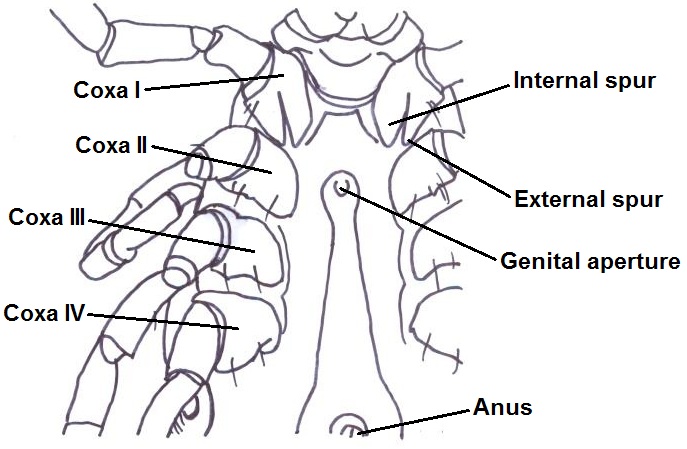
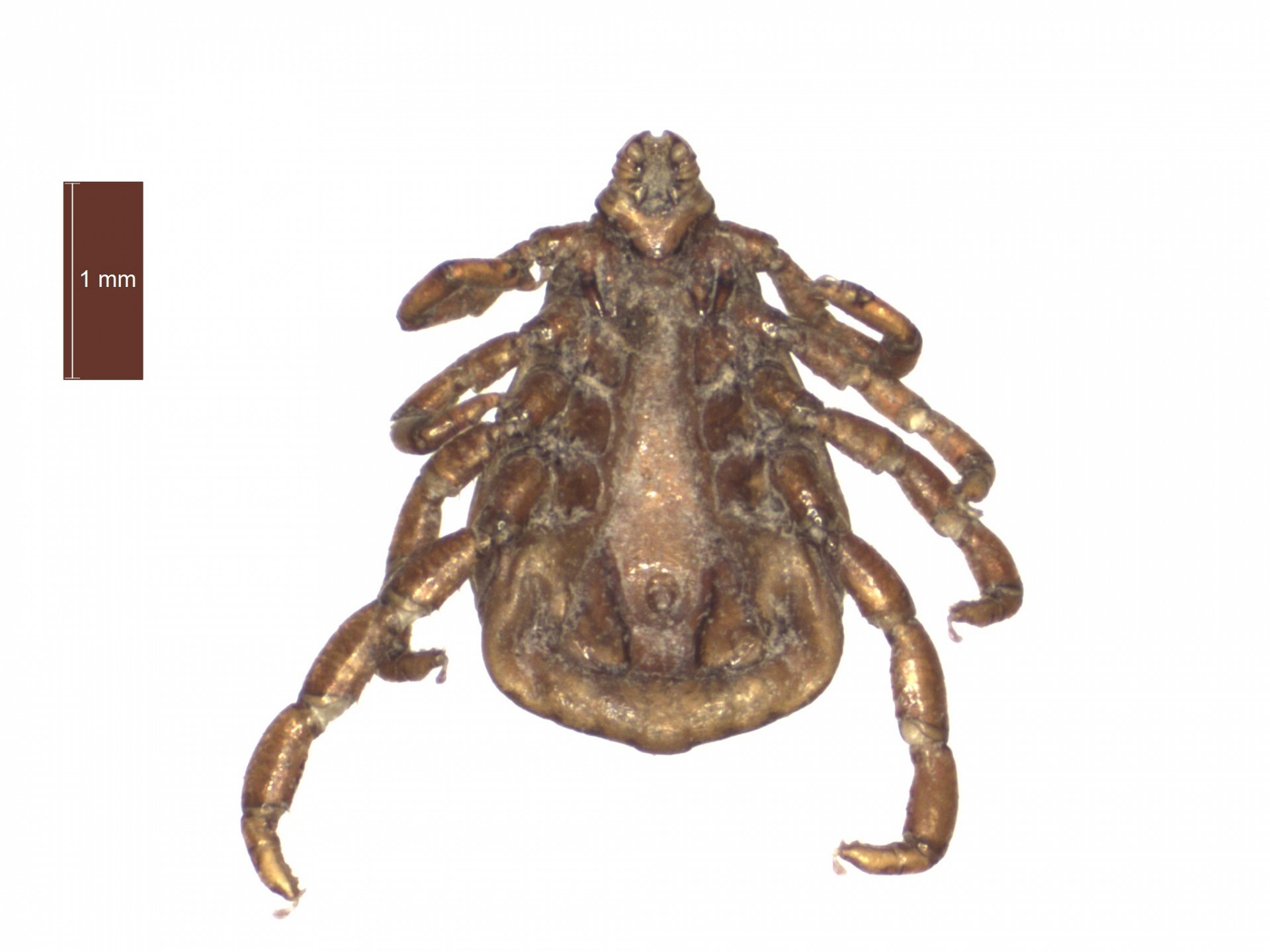
Adult female ventral features
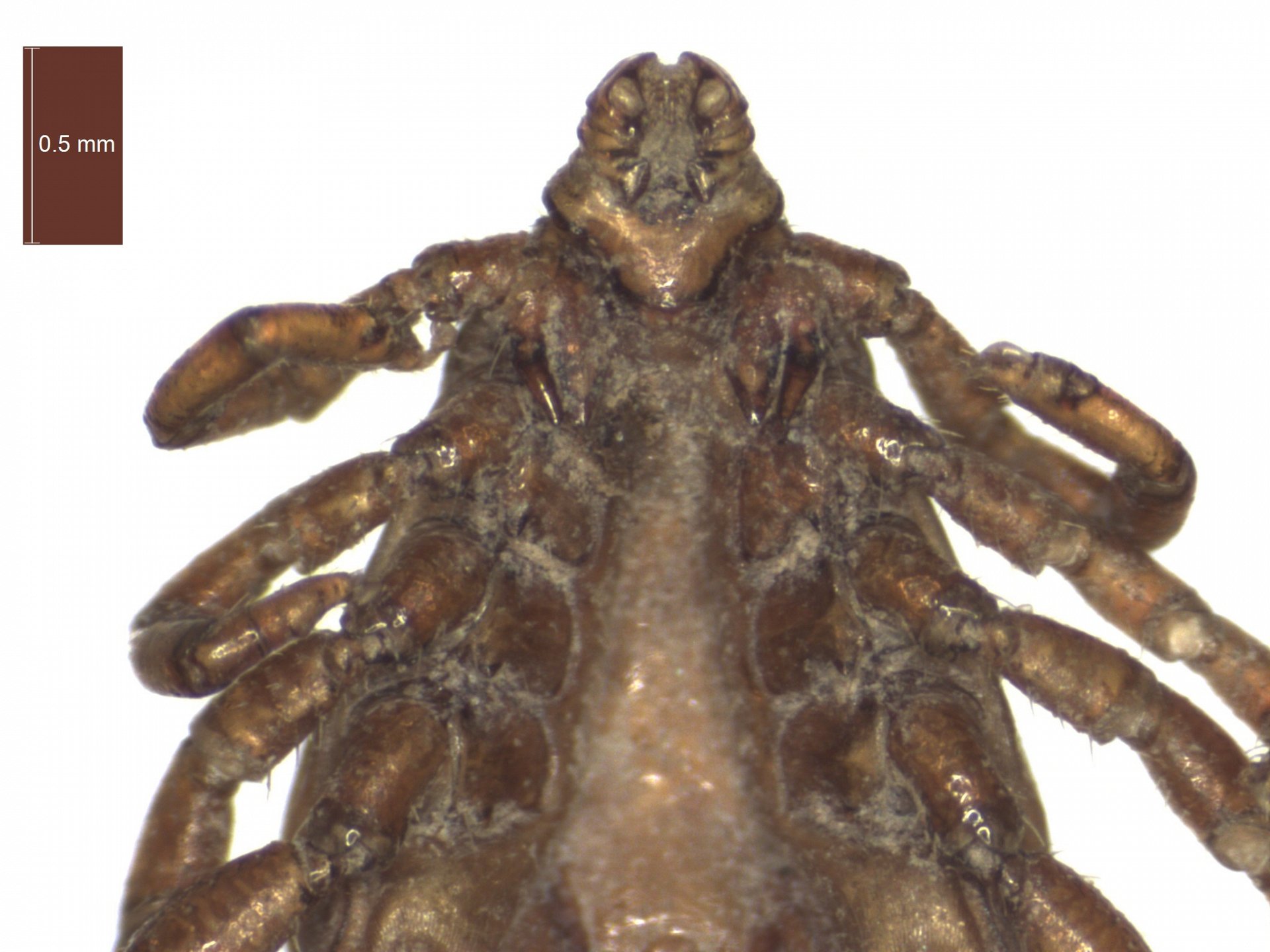
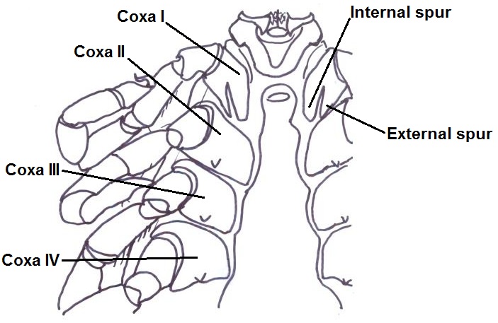
- Coxae I: large, distinct, equally paired internal and external spurs
- Coxae II-IV: small external spurs
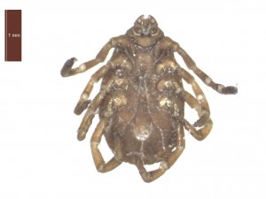
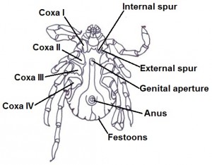
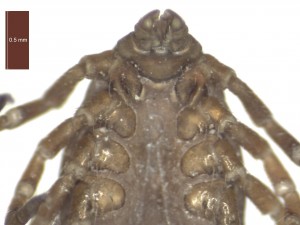
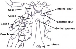
Adult female ventral view and adult female close up of coxae/ventral gnathosoma. (Click on pictures for a close up)
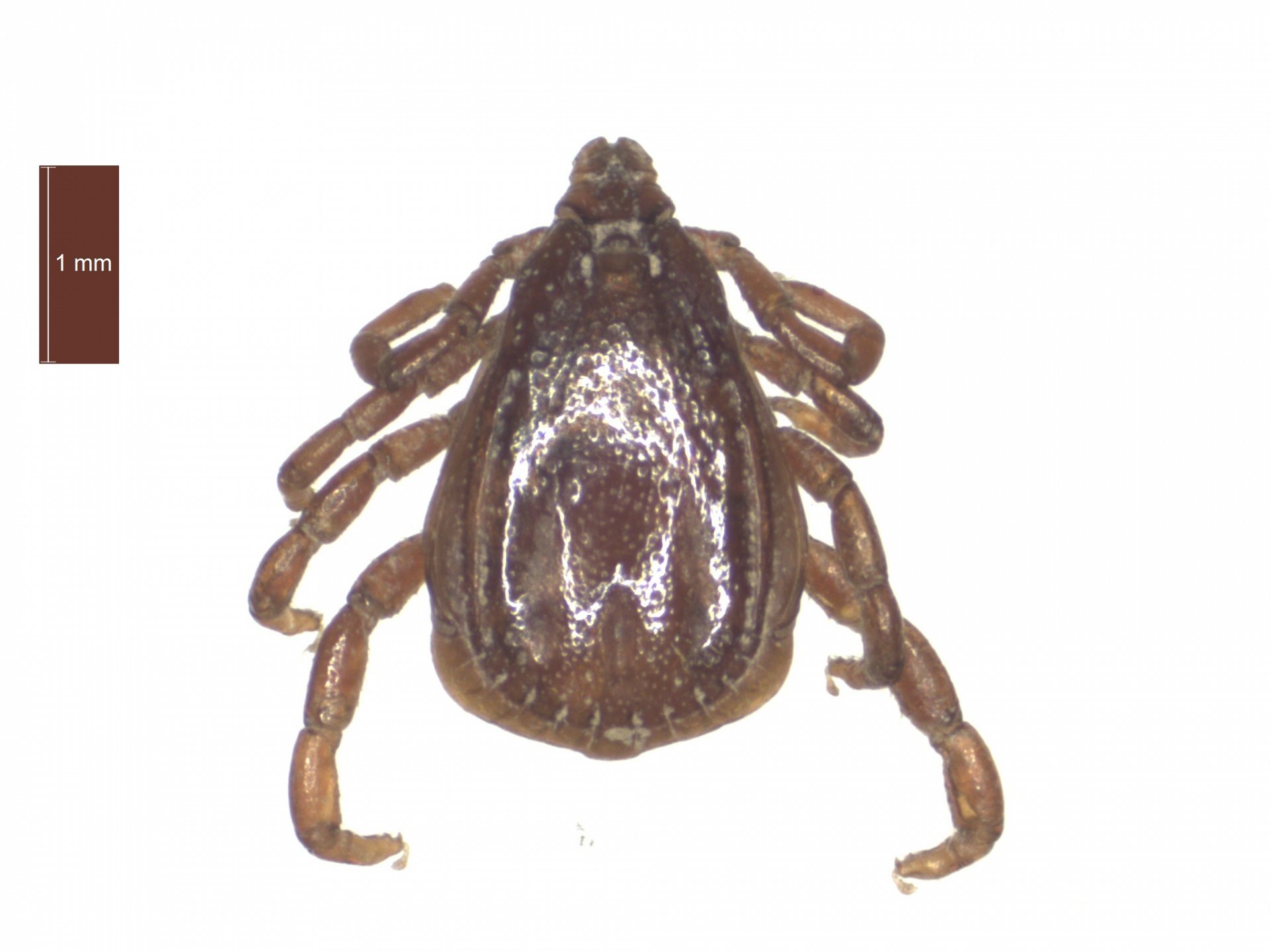
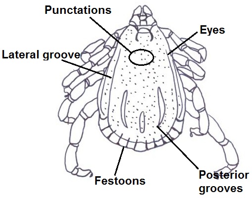
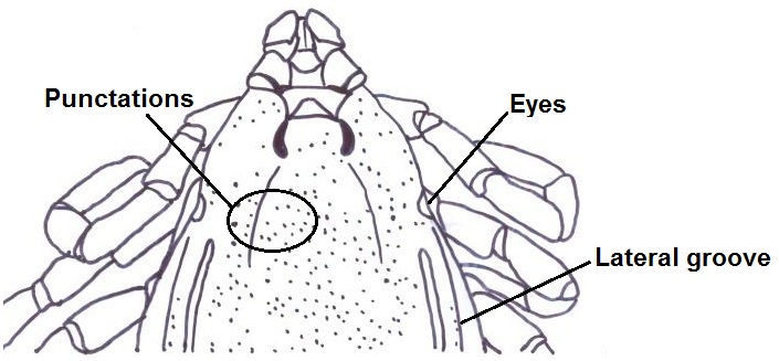
Adult male dorsal features
- Conscutum pale in colour
- Punctations are small-medium and sparse
- Lateral grooves are distinct
- Posterior grooves are distinct
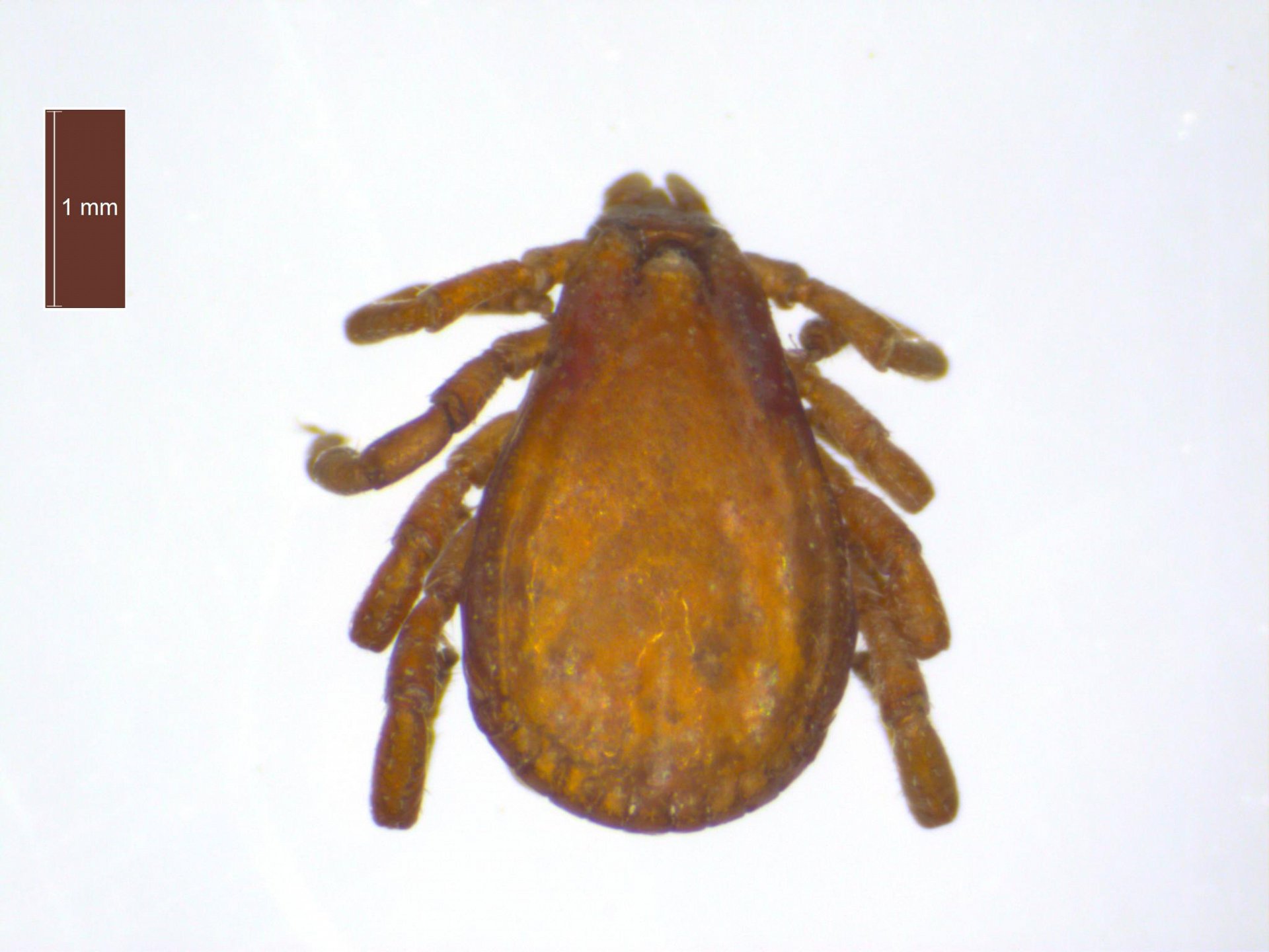
- Males measure between 3.0mm-3.8mm
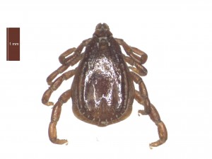
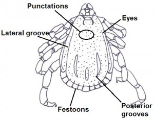
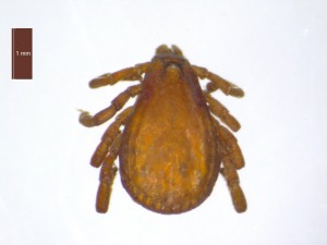
Adult male dorsal view; shape of a fed male’s body compared to an unfed male. Note: colour difference due to storage. (Click on pictures for a close up)
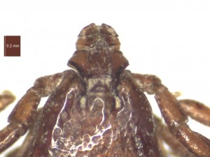
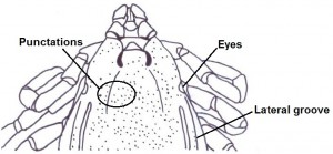
Adult male close up of gnathosoma. (Click on pictures for a close up)
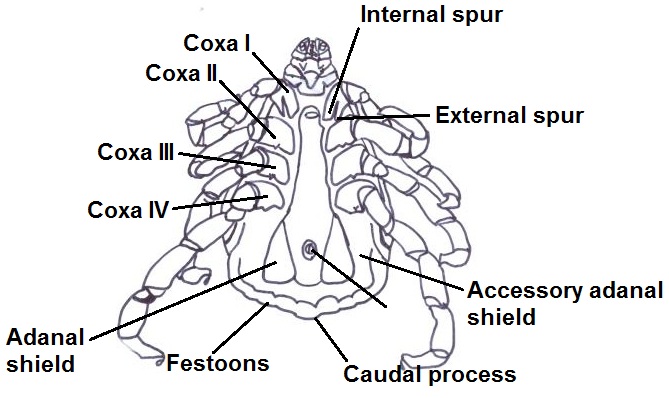
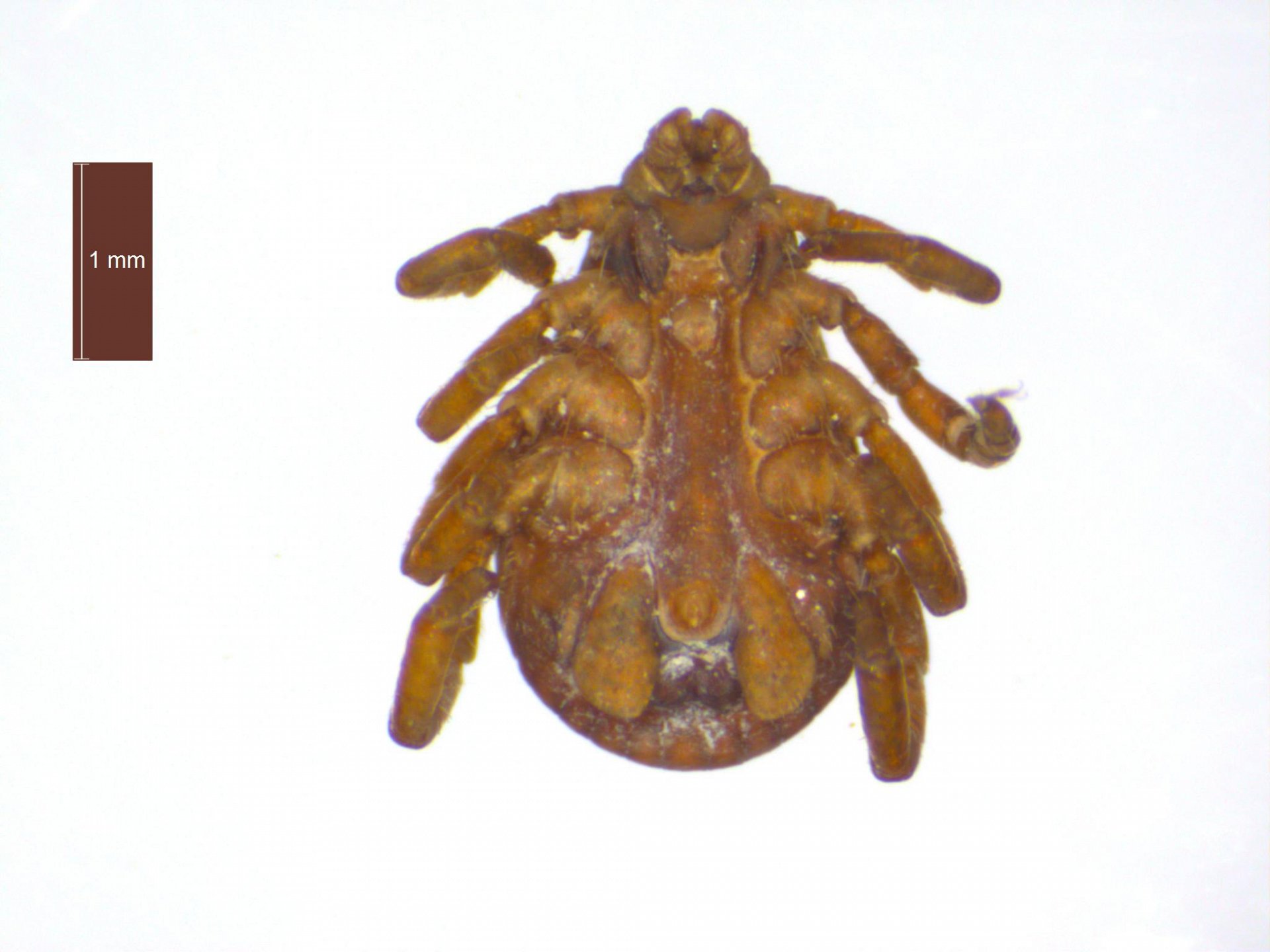
Adult male ventral features
- Coxae I: large, distinct, equally paired internal and external spurs
- Coxae II-IV: small external spurs
- Caudal process protrudes in fed males (as seen in photo above)
- Accessory adanal shields present and large
- Adanal shields present range in shape from narrow/trapezoid to broad/curve
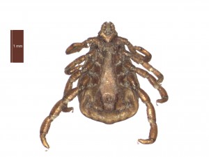
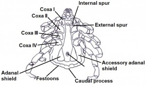
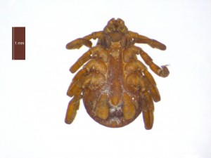
Adult male ventral view; shape of a fed male’s body compared to an unfed male. Note: colour difference due to storage (Click on pictures for a close up)

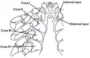
Adult male close up of coxae/ventral gnathosoma. (Click on pictures for a close up)